
A ureteric stricture is an abnormal narrowing of the ureter, the tube that carries urine from the kidney to the bladder. It can be present from birth or develop later due to ureteroscopy, injury during open or laparoscopic surgery, radiation therapy, urinary diversion procedures, or renal transplantation.
Ureteric Stricture Treatment in Pune
Dr. Rajesh Dhake > Ureteric Stricture Treatment
What Is a Ureteric Stricture?
The ureter is the thin tube that carries urine from each kidney down to the bladder. A ureteric stricture (or ureteral stricture) is an abnormal narrowing somewhere along this tube, which restricts how freely urine can flow through it. When urine can’t drain properly, it can back up toward the kidney, causing swelling, pain, and — if left untreated for long enough — a gradual decline in kidney function on the affected side.
Strictures can be present from birth (congenital) or develop later in life. In adults, they’re most often the result of scar tissue forming after an injury, infection, or a prior urological procedure.
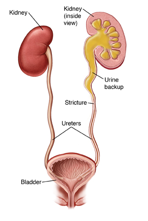
What Causes a Ureteric Stricture?
- Ureteroscopy — a stone-clearing scope procedure that occasionally leaves scar tissue behind at the treated site
- Open or laparoscopic injury — accidental injury to the ureter during pelvic or abdominal surgery for an unrelated condition
- Radiation therapy — pelvic radiotherapy can gradually cause scarring and narrowing of nearby ureteric tissue
- Urinary diversion surgery — reconstructive procedures that reroute urine flow can occasionally lead to narrowing at the new connection point
- Renal transplantation — the surgical connection made during a kidney transplant is a recognized site where strictures can form
- Prior surgery near the ureter — any pelvic or abdominal surgery close to the ureter carries some risk of scarring afterward
- Congenital narrowing — some strictures, particularly at the point where the ureter meets the kidney, are present from birth
Who Is Most at Risk?
Anyone who has undergone a ureteroscopy for a kidney or ureteric stone, or a urinary diversion procedure, carries a meaningfully higher risk of developing a ureteral stricture afterward. This is one of the main reasons follow-up imaging is recommended after these procedures, even once the original problem appears to be resolved.
Symptoms That Point to a Ureteric Stricture
- Pain in the flank or side, which may be dull or sharp
- A noticeable lump or fullness in the loin area
- Tenderness when the flank is pressed
- Recurrent or persistent urinary tract infections
- In some cases, no symptoms at all — the stricture is picked up incidentally during imaging for something else
Because symptoms can be subtle or easy to mistake for other issues, anyone with a history of kidney stones, prior ureteric surgery, or unexplained recurring flank discomfort is worth getting properly evaluated.
Treatment Options for Ureteric Stricture
Treatment depends on the length, location, and severity of the narrowing, as well as how well the affected kidney is functioning.
- Balloon Dilation — A minimally invasive endoscopic option where a small balloon is guided to the narrowed segment and inflated to stretch it open. Best suited to shorter, milder strictures.
- Endoureterotomy — An endoscopic incision made through the scarred segment to widen the passage, performed using a scope passed through the urinary tract without any external cut.
- Placement of Metal Ureteral Stents — A specialized stent is placed within the ureter to hold the narrowed segment open long-term, offering an alternative for patients who aren’t suited to repeat dilation or surgery.
- Laparoscopic Repair / Open Surgery — For longer, more complex, or recurrent strictures that don’t respond to endoscopic treatment, surgical reconstruction is used to remove the narrowed segment and rejoin healthy tissue. Where suitable, this is done laparoscopically for a smaller incision and faster recovery; more complex cases may need an open surgical approach.
Many of these procedures can be performed using minimally invasive techniques, which generally means less pain, a shorter hospital stay, and quicker recovery compared to traditional open reconstruction.
Why Early Treatment Matters
A ureteric stricture that’s left untreated doesn’t usually improve on its own — over time, the ongoing back-pressure on the kidney can lead to progressive, and sometimes irreversible, loss of kidney function. Recurrent infections are also more likely when urine can’t drain properly. Catching and treating a stricture early generally means simpler treatment options and a much better long-term outcome for the kidney.
Ureteric stricture evaluation and treatment is available at this urology clinic across Wakad, Baner, and Pimple Saudagar, open all days except Sunday. Share your details and the team will call to confirm a slot.
Frequently Asked Questions
What is a ureteric stricture, and what causes it?
What are the warning signs of a ureteric stricture?
Common signs include persistent flank pain, tenderness in the side, a noticeable lump near the flank, and recurring urinary tract infections. Some strictures cause no obvious symptoms and are only discovered incidentally during imaging done for another reason.
How is a ureteric stricture diagnosed?
Diagnosis typically starts with an ultrasound to check for kidney swelling, followed by more detailed imaging such as an intravenous urogram (IVU) or retrograde urethrogram to pinpoint the exact location and severity of the narrowing before treatment is planned.
Can a ureteric stricture be treated without major surgery?
Yes, many strictures — particularly shorter or milder ones — can be treated endoscopically through balloon dilation, endoureterotomy, or placement of a metal ureteral stent, all performed through the natural urinary passage without any external incision. Longer or more complex strictures may need laparoscopic or open surgical repair.
What happens if a ureteric stricture is left untreated?
Without treatment, the ongoing blockage can cause progressive kidney swelling and, over time, a gradual and potentially irreversible decline in kidney function on the affected side. It also increases the risk of recurring urinary infections, which is why early evaluation is strongly recommended.
What is a ureteric stricture?
A ureteric stricture is a narrowing of the ureter, the tube that carries urine from the kidney to the bladder. This narrowing can obstruct normal urine flow.
What causes ureteric stricture?
It may develop due to previous surgery, kidney stones, infections, injury, inflammation, or scar tissue formation.
What are the symptoms of ureteric stricture?
Symptoms may include pain in the side or lower back, difficulty passing urine, recurrent urinary infections, blood in the urine, or reduced kidney function.
How is ureteric stricture diagnosed?
Doctors may use ultrasound, CT scan, MRI, urine tests, blood tests, or specialised imaging procedures to identify the location and severity of the stricture.
How is ureteric stricture treated?
Treatment depends on the location, length, severity, and cause of the stricture. Options may include ureteric stenting, balloon dilation, endoscopic treatment, or reconstructive surgery.
